Picture Of Forearm Tendons ~ Forearm Tendon Anatomy Anatomy Drawing Diagram. Posted by health life media team on june 17, 2017. Tendons are fibrous cords, similar to a rope, and are made of collagen. Tendons of forearm and hand. 12 photos of the forearm tendon anatomy picture. Several joints are found between these apart from the joints and ligaments there are several muscles and tendons in the forearm.
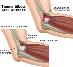
It hurts, not just when you lift or exercise, but also when you do everyday tasks, even something as basic as typing or moving the mouse on your computer. Both tendons and ligaments are dense regular connective tissue, because of its two properties: Check out our anatomy forearm art selection for the very best in unique or custom, handmade pieces from our shops. Injuries the common conditions within the tendons throughout the elbow joint comprising of the tennis elbow, and the golfer's elbow, which occur from an overuse injury to the tendons or result from. Tendinitis is an inflammation or swelling of a tendon.
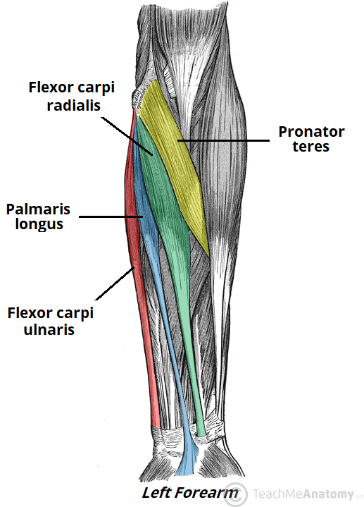
Muscles Of The Anterior Forearm Flexion Pronation Teachmeanatomy from teachmeanatomy.info This picture also contains other parts such extensor carpi radialis long, medial epicondyle of humerus, lateral epicondyle of humerus, olecranon of the ulna, extensor carpi ulnarıs, extensor dıgıtorum, flexor carpi ulnaris, extensor retinaculum, tendons of extensor digitorum and so on. Check out our anatomy forearm art selection for the very best in unique or custom, handmade pieces from our shops. Tendons are the connective tissues that connect muscle to bone. At the point when these are bothered or harmed, they end up aggravated. Both tendons and ligaments are dense regular connective tissue, because of its two properties: They have blood vessels and cells to maintain tendon health and repair injured the fcr tendon is one of two tendons that bend the wrist. It hurts, not just when you lift or exercise, but also when you do everyday tasks, even something as basic as typing or moving the mouse on your computer. In the anterior compartment, they are split into three categories:
Forearm muscle anatomy, forearm tendon pain bicep curls, forearm tendon pain from typing, forearm tendon pain from weight training, forearm tendon pain near elbow, hand tendon anatomy, shoulder tendon anatomy, wrist tendon anatomy.
Four superficial, one intermediate and three deep muscles occupy the anterior forearm. Both are made of collagen. How to treat forearm tendonitis. The forearm is the part of your arm between the wrist and elbow. Tendons are the connective tissues that connect muscle to bone. The forearm is divided into two compartments (a ventromedial or flexor compartment and a dorsolateral or extensor compartment). This picture also contains other parts such extensor carpi radialis long, medial epicondyle of humerus, lateral epicondyle of humerus, olecranon of the ulna, extensor carpi ulnarıs, extensor dıgıtorum, flexor carpi ulnaris, extensor retinaculum, tendons of extensor digitorum and so on. Forearm pain from muscle or tendon injuries can be quite debilitating. Those two tendons come from the palmaris longus muscle and the flexor carpi radialis muscle. 12 photos of the forearm tendon anatomy picture. They have blood vessels and cells to maintain tendon health and repair injured the fcr tendon is one of two tendons that bend the wrist. The two most common types of tendinitis are on the inside or outside of your elbow. (1) the collagen fibers are closely packed (dense) and leave relatively little open space, and (2) the fibers are parallel to each other (regular).
Forearm pain from muscle or tendon injuries can be quite debilitating. Forearm pain relief cause and treatment these pictures of this page are about:forearm muscles and tendons. Those two tendons come from the palmaris longus muscle and the flexor carpi radialis muscle. Injuries the common conditions within the tendons throughout the elbow joint comprising of the tennis elbow, and the golfer's elbow, which occur from an overuse injury to the tendons or result from. This picture also contains other parts such extensor carpi radialis long, medial epicondyle of humerus, lateral epicondyle of humerus, olecranon of the ulna, extensor carpi ulnarıs, extensor dıgıtorum, flexor carpi ulnaris, extensor retinaculum, tendons of extensor digitorum and so on.
Tennis Elbow Treatment Surgery Providence Orthopaedics from sgbonedoctor.com We can tell this is a ventral view of the forearm because we can see the palmar aponeurosis (a thin, tendinous sheath that is only on the palmar side of the hand) and. They have blood vessels and cells to maintain tendon health and repair injured the fcr tendon is one of two tendons that bend the wrist. Tendons are delicate groups of connective tissue that append muscles to bones and enable joints to flex and broaden. Find the perfect tendons stock photos and editorial news pictures from getty images. The two most common types of tendinitis are on the inside or outside of your elbow. Webmd's achilles tendon anatomy page provides a detailed image and description of its function as well as conditions that affect the achilles tendon. It hurts, not just when you lift or exercise, but also when you do everyday tasks, even something as basic as typing or moving the mouse on your computer. Pain, swelling, and redness of the forearm are the most commonsymptoms of the condition.
(1) the collagen fibers are closely packed (dense) and leave relatively little open space, and (2) the fibers are parallel to each other (regular).
The muscle of the common extensor tendon that is nearest this side of the arm is the extensor carpi ulnaris, which attaches to the proximal end of the fifth metacarpal, or the palm bone beneath the pinky. Check out our anatomy forearm art selection for the very best in unique or custom, handmade pieces from our shops. There are many muscles in the forearm. In most cases, conservative treatments such as avoiding any activity that. The parallel arrangement of fibers is an adaptation to the fact that. Both are made of collagen. Pain, swelling, and redness of the forearm are the most commonsymptoms of the condition. Tendons are fibrous cords, similar to a rope, and are made of collagen. A square shaped muscle found deep to the tendons of the fdp and fpl. Both tendons and ligaments are dense regular connective tissue, because of its two properties: Forearm pain relief cause and treatment these pictures of this page are about:forearm muscles and tendons. Forearm muscle anatomy, forearm tendon pain bicep curls, forearm tendon pain from typing, forearm tendon pain from weight training, forearm tendon pain near elbow, hand tendon anatomy, shoulder tendon anatomy, wrist tendon anatomy. Posted by health life media team on june 17, 2017.
The muscle of the common extensor tendon that is nearest this side of the arm is the extensor carpi ulnaris, which attaches to the proximal end of the fifth metacarpal, or the palm bone beneath the pinky. Tendons are soft bands of connective tissue that attach muscles to bones. We can tell this is a ventral view of the forearm because we can see the palmar aponeurosis (a thin, tendinous sheath that is only on the palmar side of the hand) and. At the point when these are bothered or harmed, they end up aggravated. Posted by health life media team on june 17, 2017.
The Distal Part Of Right Forearm And Hand Showing Five Tendons Of Download Scientific Diagram from www.researchgate.net It's most commonly caused by. In the anterior compartment, they are split into three categories: A tendon or sinew is a tough band of fibrous connective tissue that connects muscle to bone and is capable of withstanding tension. Check out our anatomy forearm art selection for the very best in unique or custom, handmade pieces from our shops. Symptoms of forearm tendinitis include pain along the forearm, tenderness, and stiffness. In most cases, conservative treatments such as avoiding any activity that. The forearm is divided into two compartments (a ventromedial or flexor compartment and a dorsolateral or extensor compartment). They have blood vessels and cells to maintain tendon health and repair injured the fcr tendon is one of two tendons that bend the wrist.
Pain, swelling, and redness of the forearm are the most commonsymptoms of the condition.
Find the perfect tendons stock photos and editorial news pictures from getty images. The muscle of the common extensor tendon that is nearest this side of the arm is the extensor carpi ulnaris, which attaches to the proximal end of the fifth metacarpal, or the palm bone beneath the pinky. Those two tendons come from the palmaris longus muscle and the flexor carpi radialis muscle. Both are made of collagen. The picture above is an example of a great stretch for the inner forearm muscles and tendons, do this stretch before during and after you climb both indoor and outdoor. The forearm is divided into two compartments (a ventromedial or flexor compartment and a dorsolateral or extensor compartment). Forearm tendonitis is inflammation of the tendons of the forearm. Tendons are the connective tissues that connect muscle to bone. Tendinitis is an inflammation or swelling of a tendon. Its muscle belly is in the forearm and then travels along the inside of the forearm and. We can tell this is a ventral view of the forearm because we can see the palmar aponeurosis (a thin, tendinous sheath that is only on the palmar side of the hand) and. How to treat forearm tendonitis. Webmd's achilles tendon anatomy page provides a detailed image and description of its function as well as conditions that affect the achilles tendon.
Share :
Post a Comment
for "Picture Of Forearm Tendons ~ Forearm Tendon Anatomy Anatomy Drawing Diagram"


Post a Comment for "Picture Of Forearm Tendons ~ Forearm Tendon Anatomy Anatomy Drawing Diagram"